Medical Imaging with Anti-Matter
High Resolution Positron Emission Tomography
John Sunderland
Professor of Radiology
Associate Professor of Physics & Astronomy
University of Iowa
Sponsored by PSW Science Member Michael Sandifer
About the Lecture
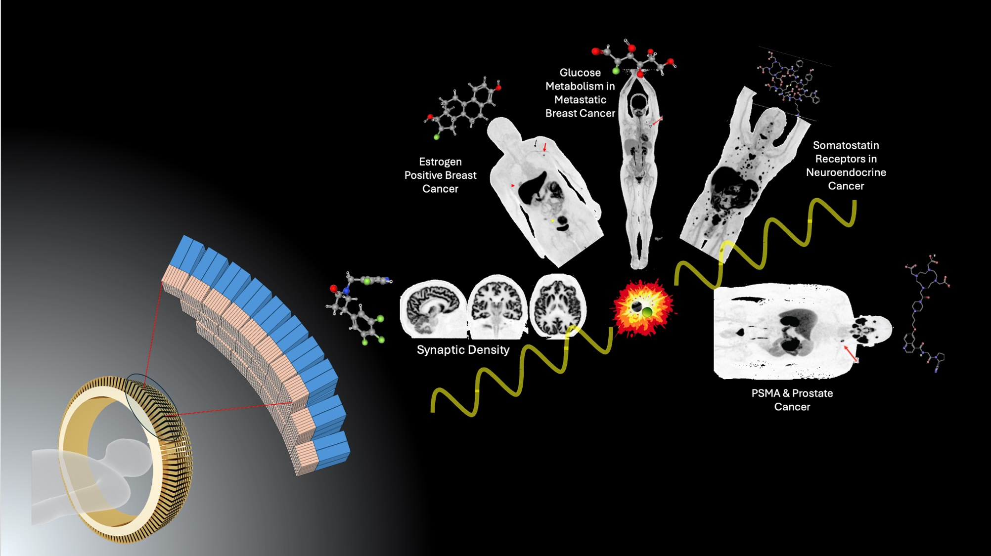
Positron Emission Tomography (PET) is a remarkably versatile clinical medical imaging modality that distinguishes itself from more well-known techniques like MRI, CT, and ultrasound imaging because PET images the underlying biochemistry of disease rather than simply the anatomy. This is critically important because most diseases are biochemical in nature, and manifest themselves first through biochemical signatures, long before anatomical changes become detectable. By directly imaging amyloid or tau protein in the brain, PET can diagnose Alzheimer’s disease in its very early stages. By imaging glucose metabolism in the body, PET can identify even tiny, hidden tumors because cancer cells rely on inefficient aerobic glycolysis to provide energy to proliferate. By imaging the expression of estrogen on the surface of breast cancer cells, PET can identify which patients will and will not respond to particular cancer therapies.
To a patient, PET imaging is relatively simple. A small injection of an unimaginably tiny amount of a radioactive drug followed by a short wait, and then a twenty minute scan of their whole body. But behind the scenes is a remarkable marriage of nuclear physics, engineering, neighborhood cyclotrons producing the necessary radioactivity, rapid radiochemistry syntheses performed under aseptic conditions, that occurred only minutes to hours beforehand. The radiopharmaceuticals that are injected were chemically engineered to be highly specific to a particular biochemical target and had to go through the rigorous FDA approval process to demonstrate safety and efficacy before becoming available.
This talk will explore the elegant but easy to understand nuclear and atomic physics that lie at the heart of PET imaging technology, and the critical role that positrons (anti-matter electrons) play in image creation. It will discuss new technological and engineering developments that continue to drive PET scanners to higher sensitivities and resolution, and new radiopharmaceutical developments that are expanding the use PET in both the research and clinical domain.
Lastly, the talk will discuss how the remarkable specificity of PET radiopharmaceuticals to bind to proteins on the surface of cancer cells has led to the exploding field of Radiopharmaceutical Therapy (RPT). By replacing the short-lived positron-emitting radionuclide on the targeting radiopharmaceutical used in PET imaging with a longer-lived beta-emitter, RPT can deliver lethal amounts of targeted radiation directly to cancer cells while minimizing damage to healthy tissue. The clinical (and commercial) success of the first of these novel radioactive drugs has resulted in a frenzied drug development environment amongst both big and small Pharma.
Selected Reading & Media References
PET Scanner Design Explained: https://www.youtube.com/watch?v=QsU3TrgArJw
Image Reconstruction: https://sites.google.com/a/fulbrightmail.org/kesnersmedicalphysics/
PET Cyclotron and Radiopharmacy Facility: https://www.youtube.com/watch?v=5aaXMLPLt20
General PET Overview: https://www.youtube.com/watch?v=l2OVu-JSU2Y
Dosimetry in Radiopharmaceutical Therapy: https://www.youtube.com/watch?v=4MLrn7Xx3Yg
About the Speaker

John J. Sunderland is Professor of Radiology in the Division of Nuclear Medicine at the University of Iowa, and of Physics & Astronomy and Radiation Oncology. In addition, he directs the Positron Emission Tomography (PET) Imaging Center and the Small Animal Imaging Core Laboratory. Previously he was Vice President for Operations at the Biomedical Research Foundation of Northwest Louisiana, directed the Creighton University Center for Metabolic Imaging and held senior leadership roles in the Society of Nuclear Medicine and Molecular Imaging.
John is widely recognized for pioneering efforts that expanded clinical PET imaging across the southern United States through centralized radiopharmaceutical production and distribution. Under the Russian Nuclear Cities Initiative, he led the development of a PET imaging center in the Urals, retraining former weapons scientists for medical applications. He was the principal author of the University of Iowa’s New Drug Application for gallium-68 edotreotide, approved by the FDA in 2019 for imaging neuroendocrine tumors—one of the few such approvals owned by an academic institution. He has also been a leading figure in advancing international PET standardization, enabling consistent quantitative imaging in multicenter clinical research.
His research spans more than four decades in medical imaging, from cyclotron targetry and radionuclide production to pharmacokinetic modeling and quantitative PET and SPECT imaging. His recent work has focused on improving the accuracy and reproducibility of quantitative imaging across different scanners and clinical sites through international harmonization initiatives. He currently co-leads the FNIH Precision Dosimetry Imaging Biomarker project, a global collaboration developing quantitative SPECT imaging standards to support radiopharmaceutical therapy trials.
John is an author on more 250 scientific publications, technical reports, and invited presentations. He has also contributed chapters to reference works in nuclear medicine and molecular imaging.
Among other honors and awards, he is a Fellow of the Society of Nuclear Medicine and Molecular Imaging and has been recognized through numerous invited lectures.
John earned a BA in Physics at Williams College, an MS and a PhD in Medical Physics at the University of Wisconsin–Madison, and an MBA at Centenary College – Frost School of Business.
Social Media Links
LinkedIn Profile: https://www.linkedin.com/in/john-sunderland-5103458/
Minutes
On November7, 2025, Members of the Society and guests joined the speaker for a reception and dinner at 5:45 p.m. in the Members’ Dining Room at the Cosmos Club. Thereafter they joined other attendees in the Powell Auditorium for the lecture proceedings. In the Powell Auditorium of the Cosmos Club in Washington, D.C., President Larry Millstein called the lecture portion of the 2,524th meeting of the Society to order at 8:02 p.m. ET. He began by welcoming attendees, thanking sponsors for their support, announcing new members, and inviting guests to join the society. Scott Mathews then read the minutes of the previous meeting which included the lecture by Neil Johnson, titled “AI’s Jekyll-and-Hyde Tipping Point: The Science of When Good AI Will Go Bad”. The minutes were approved as read.
President Millstein then introduced the speaker for the evening, John Sunderland, of the University of Iowa. His lecture was titled “Medical Imaging with Anti-Matter: High Resolution Positron Emission Tomography”.
The speaker began by describing positron emission tomography (or PET) as being inherently multidisciplinary, involving physics, biochemistry, synthetic organic chemistry, medicine, and engineering. He indicated that PET does not image anatomy, rather it images specific biochemistry in the human body. He presented an example image, showing the glucose metabolism in a patient, after injecting a tiny amount of radioactive glucose. He described the “beta-plus” decay process, in which a nucleus with too many positive charges emits a positron (the anti-matter version of an electron). He showed how the positron combines with an electron, after propagating a very short distance, and creates two gamma rays of exactly 511 keV, propagating in opposite directions. Sunderland described three discrimination processes, used to separate electron-positron annihilation from other detection events. These included energy discrimination, temporal discrimination, and geometrical discrimination. Energy discrimination, or pulse height discrimination, relies on the fact that the magnitude of the electrical pulse generated at the detector is proportional to the energy of the detected particle, and electron-positron annihilation generates monoenergetic gamma rays. Temporal discrimination, or coincidence counting, relies on the fact that the two gamma rays arrive at the detectors almost simultaneously (within a few nanoseconds). And geometrical discrimination, which relies on the fact that the two gamma rays propagate exactly 180-degrees from one another, meaning that they are detected on diametrically opposed detectors. These three discrimination mechanisms result in exquisite sensitivity to electron-positron annihilation, filtering out all other detection events, and allowing practitioners to use extremely small quantities of radioactive materials.
The speaker discussed the motivation for characterizing and imaging glucose metabolism in the human body, saying that most cancer cells metabolize glucose extremely inefficiently. As a result, cancer cells need to burn glucose at a much higher rate than normal cells, in order to support cellular function. Sunderland showed a PET image of a patient with breast cancer. He said that although the patients CT scan came back normal, four malignant lymph nodes were clearly visible in the PET image.
Sunderland discussed the fact that PET scanning is a “Tracer methodology”; that is to say that the quantity of injected material must be so small as to not perturb the biochemical system being probed. He discussed the biochemical optimization of the tracer molecules used in PET scans, giving the example of labeling glucose with carbon-11, as compared to fluorine-18. He said that C-11 glucose would be metabolized into CO2, and would rapidly begin to diffuse throughout the body. Fluorodeoxyglucose, labelled with F-18, is metabolized differently and therefore remains sequestered in the cells.
The speaker presented a brief introduction to nuclear physics, emphasizing radioactive isotopes. He showed the chart of the nuclides, describing beta decay: neutron-rich nuclei decaying with the emission of an electron, and proton-rich nuclei decaying with the emission of a positron. He mentioned nuclei which decay by alpha emission, saying they were relevant to radiotherapy. Sunderland said that many unstable nuclei, whether decaying by alpha or beta processes, will create a nucleus in an excited state, which subsequently emits a gamma ray. He indicated that gamma rays, produced by nuclear decay or electron-positron annihilation, are important “messengers”, as they have a high likelihood of leaving the body and being detected in scintillators.
The speaker discussed processes for producing PET-scan tracers, including: using cyclotrons to create the isotopes, chemical synthesis under aseptic conditions, and remote handling in “hot cells”, due to the high levels of radioactivity. He discussed the desired characteristics for these isotopes: half-life long enough to deliver the isotopes to a clinical site, but short enough to allow image formation in a reasonable period of time. Sunderland discussed the diversity of PET radiopharmaceuticals, including about 15 that are approved for clinical use. He presented a list of the most common compounds, the isotopes they incorporate, and the specific clinical uses of each. He showed specific examples of PET imaging for prostate cancer, breast cancer, neuroendocrine tumors, and amyloid proteins for Alzheimer’s disease. Sunderland claimed that newer compounds currently under development are likely to have a significant impact on a wide variety of disease conditions, including: colorectal cancer, pancreatic cancer, ovarian cancer, cardiac disease, neurological disease, T-cell activation, etc.
The speaker then described the functioning of a PET scanner. He described the detector array; rings of tens of thousands of scintillation crystals, optically coupled to light sensors, feeding signals to fast electronics which implement discrimination and recording of each event. He indicated that older PET systems use photomultiplier tubes for light detection, but that modern systems use silicon photomultiplier arrays, which dramatically increase the image quality and resolution. He described the computer-based image reconstruction process known as tomography: the numeric reconstruction of a 3-dimensional image from a series of 2-dimensional “slices”, each acquired from a different perspective.
The speaker ended his talk by describing “targeted radiopharmaceutical therapy”: injecting much larger doses of radioactive compounds, which will preferentially bind to tumor cells, such that the tumor cells receive a much higher percentage of the radiation than the surrounding healthy tissues. He described how PET imaging techniques could be used to measure the absorbed dose of radiation in specific organs, in order to “personalize” the dosage for each patient.
The lecture was followed by a Question and Answer session.
A member asked about the scintillators currently used in PET imaging, asking if NaI (sodium iodide) crystals were still used. Sunderland responded that NaI is still widely used for “gamma cameras”, but that most modern PET systems employ lutetium orthosilicate (Lu2SiO5) crystals, which are very fast, very dense, and have good energy resolution.
A new member asked about “time-of-flight” imaging and whether such techniques could be pushed to the femtosecond scale. Sunderland responded that there are physical limits, primarily associated with the size of the scintillation crystals; i.e. the time delay associated with a gamma ray interacting with the front surface, the back surface, or somewhere in between. Decreasing the thickness of the scintillators would reduce the uncertainty in time, but would compromise absorption.
A guest on the live stream asked if it would be possible to combine data from PET scans, CAT scans, MRI images, and X-ray images, and use AI to improve diagnoses and treatment. Sunderland responded “theoretically, absolutely ‘yes’”. However, he indicated that the primary difficulty of such “data fusion” is HIPAA (Health Insurance Portability and Accountability Act), which limits the ability of practitioners to share medical data.
After the question and answer period, President Millstein thanked the speaker and presented him with a PSW rosette, a signed copy of the announcement of his talk, and a signed copy of Volume 17 of the PSW Bulletin. He then announced speakers of up-coming lectures and made a number of housekeeping announcements. He adjourned the 2,524th meeting of the society at 9:50 pm ET.
Temperature in Washington, DC: 15.6° Celsius
Weather: Mostly cloudy
Audience in Powell auditorium: 66
Viewers on the live stream: 32
For a total of 98 viewers
Views of the video in the first two weeks: 183
Respectfully submitted, Scott Mathews: Recording Secretary