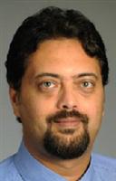
Advances in Medical Imaging
Ahmed M. Gharib
Radiologist
National Institutes of Health
About the Lecture
With the development of the first X-ray image in 1895 the advancement of medical imaging began. Since then, these advances have achieved huge leaps which allowed for the better understanding and management of different diseases. This presentation will focus on Coronary CT and MR at high magnetic field as a venue to discuss some of the advances in medical imaging and the ability to overcome the challenges of imaging these, small yet critical, vessels. The lecture will also discuss the current and potential applications of imaging technology in improving our understanding of atherosclerosis.
About the Speaker

AHMED M. GHARIB has been a Staff Radiologist at the NIH since 2005. His clinical interests are in multimodality cardiovascular imaging (including cardiac CT and MR) and body MRI, while his research efforts are focused on early detection of atherosclerotic plaque. He serves as a primary and associate investigator on several research protocols. His research appointment is in the National Institute of Diabetes Digestive and Kidney Diseases (NIDDK) for his research work as the senior radiologist in the intramural research laboratory of the National Institute of Biomedical Imaging and Bioengineering. He also serves as a clinician in the Diagnostic Radiology Department. Before coming to NIH. He served as a Chief Resident at the University of Washington, in Seattle. He is boarded in both Diagnostic Radiology by the American Board of Radiology and in Nuclear Medicine by the American Board of Nuclear Medicine. His fellowships include Chest Radiography (University of Washington), Cross Sectional Imaging (Johns Hopkins University) and Molecular Imaging (NIH). He currently serves as a referee for multiple major imaging journals including Radiology, the American Journal of Radiology, Investigative Radiology and Magnetic Resonance in Medicine. He has also been an invited speaker at multiple national and international meetings. He received his M.B., Ch.B. (Bachelor of Medicine and Surgery) degree from the Faculty of Medicine, the University of Alexandria, Egypt, in November 1993. He served an internship in Internal Medicine at the University of Washington; a residency in Nuclear Medicine and Nuclear Cardiology at the University of Washington; and a residency in Diagnostic Radiology at the University of Louisville.
Minutes
President Larry Millstein called the 2,257th meeting to order at 8:16 pm September 25, 2009 in the Powell Auditorium of the Cosmos Club. The minutes of the 2,256th meeting were provisionally approved after a short discussion.
Mr. Millstein introduced several new members of the Society and then introduced the speaker of the evening, Mr. Amed M. Gharib of the National Institutes of Health. Mr. Gharib spoke on “Advances in Medical Imaging.”
Mr. Gharib asked the lights be reduced, and noted that people in imaging like to sit in the dark. Low light, he said, increases contrast, and contrast is very important. After we got the light down, the sound up, and the pictures of Mr. Gharib’s kids off the screen, the discussion began.
He started with CT, computed tomography. X-rays are absorbed at different rates by different types of tissues, and the difference between in-radiation and out-radiation through all the locations in the body are digitized, 1 for water, 1000 for bone, and so on, and the data are used to compute and graph images of the kind of tissue at each location. It requires a tube rotating around the patient shooting rays through. Contrast materials are also put into the body. This kind of imaging has been around for 20 years. It is still very useful for looking at anything large.
He showed an example of an image of a lung which had a little nick in it. That, he said, was where a tumor was removed. With CT 3D imaging, it was possible to remove it without opening the chest.
Important recent developments are in imaging things that are very small. An example is imaging the coronary arteries, small arteries that feed the heart tissue.
The existing procedure involves putting a catheter into a large blood vessel in the groin area, pushing it up to the heart, and then injecting a contrast material with an X-ray tube on. This is done repeatedly in the small arteries while recording data. This enables imaging of the small vessels that feed the heart itself. He showed an example of using this technique to show a small but serious aneurism over an aorta. Resolution down to .41 mm is used to get the needed detail in the pictures. The catheter can also have a tool on it to use to open up clogged arteries.
Imaging an artery this way does have risks and difficulties. The heart is jumping at 60 to 90 beats a minute and the arteries are small. He said it is like imaging a spaghetti on a trampoline. They slow the heart down to improve the picture, shoot with the highest resolution, and use a fast shutter speed. They also have increased the number of detectors. At first they used only one, then two, then four, eight, and 16, and now they are up to 320 detectors all running at once. They average over heartbeats, and the more detectors they have, the fewer heartbeats it takes to get an image. They also time the shot for the 100 to 200 ms when the heart is relaxed and relatively still. All this reduces the amount of X-ray energy needed.
Until recently, it took a 180-degree rotation of the tube to get a set of pictures. A simple innovation, use of two X-ray sources, reduced that to a 90-degree rotation. Coupling that with the other improvements, they can get a picture in a single heartbeat. This improves the picture by reducing the total amount of movement that needs to be factored out of the picture. It also reduces the radiation exposure.
Plaque in the heart vessels is either calcified or not calcified. The calcified plaque is plaque that is healed, but the plaques pop off and block vessels, that is, a heart attack.
He discussed his handyman, who is in his 60's and was apparently healthy, and who offered to let Mr. Gharib scan him. Mr. Gharib did so and found serious restrictions in his heart vessels, critical heart disease, although he had no symptoms, even during a stress test. The restrictions were in areas of soft plaque, not the calcified plaque. This pattern is much like that of the recently deceased Tim Russert. Mr. Gharib’s handiman received a stent, unlike Mr. Russert.
The coronary artery runs in the muscle. When muscle contracts, the artery contracts. Pictures of the heart during the contraction, made possible by the reduced imaging time, show the amount of restriction during the contraction.
He showed a scan of a patient with a congenital shunt. It looked like a dirty pipe. They were able to go in and clean it up.
The new techniques have revealed many anomalies in the heart tissue, many of them congenital. One of them is the Job syndrome, named for the miserable, patient Job described in the Bible. There are many symptoms, but the main one is that they get many infections. Interestingly, they do not get atherosclerosis, but they have deformed, wobbly, wiggly blood vessels in the heart.
He mentioned “knockout mice,” mice bred with a missing gene to study the effects. Now they have “knockdown humans,” humans with missing genes and resultant anomalies. It is a mutation called the “Stat 3" mutation that causes Job syndrome.
He next discussed something he called “almost miraculous,” magnetic resonance imaging (MRI). He seemed really enthused about getting images with no radiation at all, as it enables him to put people in the scanner freely and indefinitely. The body is put in a magnetic field; a radio frequency signal is sent in; the signal is altered by the magnetic resonance to different degrees by different body structures; the altered outcoming signals are digitized and images are computed in the same way as in CT. Since the picture is computed, image elements can be selected and located to graph any slice of the tissue they may need.
This works well with large vessels. With small vessels, it works pretty well also if they inject some contrast material. In some ways, it works better than CT.
He gives his patients beta blockers to slow down their heartbeats. He said when he does that, he has to give himself one, too, to improve his patience and timing. Because of the heart’s [annoying] beating, the image data must be gathered in a very short time period. Timing is critical; if the heart is not “smiling,” the image is ugly.
They discovered, with these improved image techniques, a phenomenon called “small heart attacks,” indicated by small white dots in the heart tissue. These small heart attacks have no symptoms, but they are a newly identified phenomenon. Another new phenomenon: where macrophages occur, there is angiogenesis, growth of new tissue. There new vessels are formed, and sometimes poorly formed, and they leak.
A great advantage of the new techniques is the absence of potentially harmful aspects. High resolution, quick imaging enables them to get images with little beta blockers, little or no contrast material, and no radiation. This frees them to make all the images they want. Mr. Gharib’s enthusiasm about this prospect was evident. It was this liberty that encouraged him to do the scan of his handyman, who had no symptoms.
After the talk, the first question for the speaker was, with all those anomalies, how many people are normal. He didn’t know. He guessed perhaps 50% of us in the room have some atherosclerosis.
He clarified that the 320 detectors used in imaging now are for coverage, not resolution. They increase the area or volume that can be covered in one sweep.
A young woman asked about people with tattoos. Are the horror stories she has heard true? They are, he said. People don’t always feel much pain, but they sometimes do. Someone said the problem is most severe with black inks, which contain iron.
He observed that certain tedious and uncomfortable scanning processes work better in certain Asian cultures, where people are more accepting, obedient, and disciplined. Totalitarianism has its merits.
After the talk, Mr. Weinstein presented a plaque commemorating the occasion. He made the parking announcement. He announced the next meeting. He invited suggestions of future speakers. He encouraged membership, participation, and sponsorship. Our newest member, Mr. Gharib, offered free scans to any other members. Finally, at 9:45 pm, Mr. Weinstein adjourned the 2,257th meeting to the social hour.
Attendance: 52
The weather: Cloudy, moist
The temperature: 18°C
Respectfully submitted,
Ronald O. Hietala,
Recording secretary