Multiphoton Imaging Excitation Microscopy
Subcellular Imaging In Vivo
Robert S. Balaban
Scientific Director
National Heart Lung and Blood Institute
National Institutes of Health
About the Lecture
Recent developments in the use of non-linear optical techniques have provided the unique ability to image the molecular structure and function of cells in vivo. This technology is on the verge of bringing together cell biology, physiology and medicine in intact systems where sub-cellular events can be observed in the context of the intact body. The basic use of two-photon excitation fluorescence microscopy essentially permits the delivery of visible photons deep into tissues using infrared light making intra-vital fluorescence microscopy feasible and practical. New optical excitation and detection schemes for the microscope are resulting in several fold improvements in the signal to noise characteristics of this approach providing the most sensitive method of imaging any optical fluorescence probe. This lecture will review an example using a second harmonic imaging method to image the macromolecular structure of the arterial wall that has led to a new theory of early arteriosclerosis development.
About the Speaker

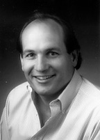
ROBERT S. BALABAN is Scientific Director of the National Heart, Lung, and Blood Institute (NHLBI) at the National Institutes of Health. He is also Chief of the Laboratory of Cardiac Energetics, Division of Intramural Research, NHLBI. He attended the University of Miami as an undergraduate in marine biology and received Bachelor of Sciences degrees in Biology and Chemistry in 1975. He attended graduate school at Duke University with an NIH training fellowship where he received his Ph.D. in Physiology and Pharmacology in 1980. The title of his dissertation was “The coupling of aerobic metabolism to active ion transport in the kidney”. He was awarded a NATO Post-Doctoral Fellowship in the Department of Biochemistry at The University of Oxford (1980-1981). He joined the NHLBI as a staff fellow in the Laboratory of Kidney and Electrolyte Metabolism in 1981. After continuing his work in renal energetics and the discovery of functional organic osmolytes in the kidney, he redirected his efforts to the study of the heart. In 1988 be became the Chief of the newly formed Laboratory of Cardiac Energetics in the NHLBI. He was appointed to Scientific Director of the Laboratory Research Program in December of 1999. He became the overall Scientific Director of the NHLBI intramural program in 2004. His primary research interest is in the overall energy economy of intact biological tissues with a recent focus on the function of the heart. Due to his interest in the function of intact systems, his research has relied heavily on the development and use of non-invasive technologies. These technologies include magnetic resonance imaging and spectroscopy, optical imaging and spectroscopy, positron emission tomography and ultrasound approaches. He has published over 250 papers in peer-reviewed journals on a wide variety of research topics. He is the co-inventor on 10 patents on technology developed for these studies. He has served as President of the International Society for Magnetic Resonance in Medicine and President of the Society for Cardiovascular Magnetic Resonance. He is also a member of The American Physiological Society, The American Society for Cell Biology, and The Biophysical Society.
Minutes
President Larry Millstein called the 2,252nd meeting to order at 8:23 pm March 6, 2009 in the Powell Auditorium of the Cosmos Club. The minutes of the 2,251st meeting were read and approved.
Mr. Millstein introduced the speaker of the evening, Mr. Robert S. Balaban of the National Heart, Lung, and Blood Institute. Mr. Balaban spoke on “Multiphoton Imaging Excitation Microscopy: Subcellular Imaging In Vivo.”
Mr. Balaban began by saying that there is an emerging trend in systems biology as a result of improved imaging. “We are on the verge,” he said, of “putting the system and the body back together ... [to] find out how it all works.” This started with gene sequence and gene expression.
Now we can also look at proteins and protein expression. Many things were known from the old “grind and find” biochemistry, such as the mechanism of diabetes. Now, he said, he believes it, because he has actually seen what has happened when he gave an animal insulin. We, the biologist, the physicist, the physician, can now start doing experiments. Imaging is going to be the platform for interface among the professions.
He described two-photon excitation fluorescence microscopy. In conventional microscopy, it is very difficult to get blue light into tissue. He showed a two-photon Jablonski energy diagram to show how this works. They use a 360 nanometer light; at the focal point, the two co-act and deliver blue light deep in the tissue. As good as this is, multiphoton excitation fluorescence is much better. The latter yields a depth of penetration 10 to 100 times that of single photon excitation. It has other advantages, also, including limited photo-damage and bleaching and enhanced contrast.
This, he said, is one of their tricks, one among many.
Another trick is gathering more light with a parabolic mirror around the aperture of the objective. This could theoretically yield nine times as much light; in fact it yields five times. This enables them to do experiments 25 times faster because of the much clearer images resulting from the increase in light from the area of interest.
He told of adapting a method of correcting for the atmosphere, developed for astronomy in the space program. It is done by bouncing light off a mirror on the moon. The process is called adaptive optics. In Mr. Balaban’s mode of microscopy, the scattering comes from the tissue rather than the atmosphere. The tissue surrounding the subject area becomes one of the lens elements using this technique, once it became determinable how the light was being scattered by the tissue.
Using these methods of microscopy, researchers are able to get very fine, actual pictures of objects the size of mitochondria. The structures of arterial walls have been displayed in fine detail.
Until recently, it was not possible to get photons into and back out of a live heart. There is a layer of tissue called the visceral pericardium elastin. It has two types of elements, one elastic and one muscular, which are always at a fixed angle to each other. Studies of the structure with these microscopy techniques revealed that this fixed angle gives the heart wall its elasticity.
Much has been learned about cardiovascular conditions with photon excitation microscopy. Mr. Balaban showed illustrations of the stages of atherosclerosis. The advanced stages do look ugly, with the plaques, the distortion of tissue, and ruptures of the vessel walls making the damage very obvious.
One of the questions about atherosclerosis is: Why do plaques form near the arterial branches? He said there is no turbulence there. One possibility is that collagen is exposed at the vessel branches. He said this is likely a mechanical requirement, collagen being involved in the elasticity of the vessel wall. This exposure of collagen may provide the electrochemical basis for the beginning of the deposit of low density lipoprotein (LDL), often called “bad cholesterol.” He showed remarkably clear pictures of the collagen sites and how LDL deposit progresses at those sites. Researchers have also determined that LDL binding at those sites is non-linear, consistent with the finding that small changes in plasma are associated with large clinical effects. Also, this kind of imaging is capable of rendering in three dimensions. Vessel blockages that are invisible in two dimensions are shown clearly in three dimensions.
He drew an analogy from computer engineering, beginning with the question: How do you analyze complex systems? You don’t take them apart. If you do that, they stop working, and there is no way to infer the whole system from the parts. A computer engineer told him that you analyze a board by measuring what happens at the check points that the designers built into the board. This sent him on a search for such check points in the biological systems he studies.
One such point is the NADH (nicotinamide adenine dinucleotide plus hydrogen) found at cell walls. It is useful because it indicates the energy potential available for work in the cell. He said it is analogous to voltage. He described how it is imaged using NAD(P)H fluorescence, which emanates primarily from the NADH bound to the site.
He described other methods of image refinement, including:
retrospective motion compensation
image averaging
deformation correction
slice tracking
focus control built into the microscope
motion simulated from tracking data
Finally, Mr. Balaban closed by discussing how these fine views of microscopic elements of tissue are giving us a much better macro view of the biological whole.
In the question period, someone asked how he got multicolor images from monochromatic light. That is just coding, he said. They put different colors on different views just to make the differences easier to see.
In answer to another question, he said they can produce images that exceed the wave length of light by only three times.
Someone asked, given his explanation of why plaques develop, why does it take 20 years for clots to form. He discussed the formation of clots in more detail, but admitted that the answer to that question is not known. What he told us on that, he said, is “semi-science.” He mentioned that many strokes are just clots that break off, flow through the channel, and block the blood stream when they reach a smaller channel.
Responding to a question involving evolution and atherosclerosis, he asked: Is there any evolutionary value to the survival of a man over 50? If he were to die right now, he said, his kids would do okay. So there has been no evolutionary pressure on atherosclerosis. He speculated that he, needing glasses and having had five knee operations, probably would not be alive had nature taken her course. He would have been caught by a coyote.
After the talk, Mr. Millstein thanked our speaker and presented a plaque commemorating the occasion. He announced the next meeting. He made the parking announcement. He invited suggestions for speakers and encouraged guests to consider joining. Finally, 10:06 pm, he adjourned the 2,252nd meeting to the social hour.
Attendance: 52
The weather: Cool and mild
The temperature: 12°C
Respectfully submitted,
Ronald O. Hietala,
Recording secretary